

Physical Examination
Since a varicocele cannot always be seen, a physical examination of your testicles in standing or lying positions may be required. If the physical exam proves to be ineffective in detecting the enlarged veins, then the doctor may have to perform a scrotal ultrasound in order to get a better look inside the scrotum. Typically by the time patients come to our office they have already been diagnosed with a varicocele.
Scrotal Ultrasound
Ultrasound uses high-frequency sound waves to create an image inside your body. Since the scrotum is located on the outside of the body this is an ideal test. What does the ultrasound look for? The ultrasound is done using a maneuver called “Valsalva.” The technologist may have you bear down like you’re going to the bathroom to see if the veins enlarge. If the veins meet a certain size criterion then the diagnosis of a varicocele can be made.1 Sometimes the abnormal flow can also be visualized. If you do not have an ultrasound our specialist will either order one at a location close to you or perform this study at our office; our doctor is dual board certified and can perform ultrasound studies himself.
CT Scan
A CT uses x-rays to look inside the body to provide detailed anatomy. Sometimes there can be other processes going on inside the abdomen that may block the flow in the testicular vein. The CT scan also performed to accurately measure the size of the testicular veins. Our specialist routinely orders this exam prior to treatment to allow for more precise placement of the coils to prevent migration or complication.
Request an Appointment
Please note that although we strive to protect and secure our online communications, and use the security measures detailed in our Privacy Policy to protect your information, no data transmitted over the Internet can be guaranteed to be completely secure and no security measures are perfect or impenetrable. If you would like to transmit sensitive information to us, please contact us, without including the sensitive information, to arrange a more secure means of communication. By submitting this form you consent to receive text messages from CVI at the number provided. Msg & data rates may apply. Msg frequency varies. Unsubscribe at any time by replying STOP.
What is Varicocele Embolization?
Varicocele can be treated by an embolization procedure or surgery. Embolization is a non-surgical, outpatient, minimally invasive technique that uses x-ray guidance to place tiny coils and embolic fluid in the abnormal blood vessels causing them to close down.
Am I a Candidate for Varicocele Embolization?
Treatment is offered for males who have:
- Fertility problems (problems fathering a child)
- Pain
- Boys with a smaller testicle; at a higher risk for fertility problems when they are older
How Is Non-Surgical Embolization Performed?
- You will receive twilight sedation to help you relax
- Our specialist numbs the skin on the neck or groin
- A tiny catheter is placed
- Foam and small coil plugs placed into the vein
- The catheter is removed
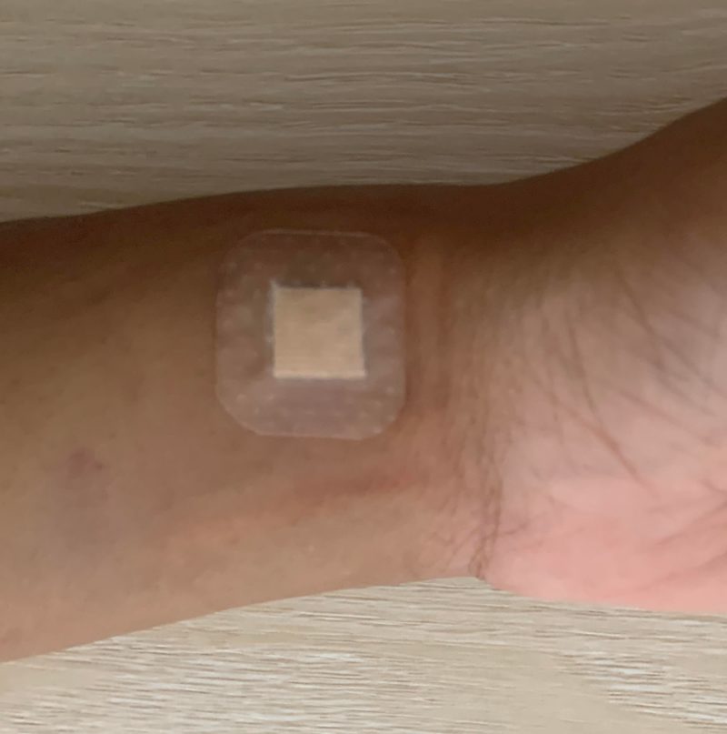
- Band-Aid is applied

We are Here to Help
Request an Appointment to meet with our varicocele specialist who will review your imaging, labs and history to determine if you are candidate for the procedure, and the outcomes you can expect. Each person is an individual and should discuss the potential risks and benefits of embolization and other treatments with our doctor to decide which option is best.
Appointments are available via an online video telehealth platform or in person at one of the offices in Los Angeles, Orange County or San Diego. Why should you choose us? Read here
1.) Cimador, M, Di Pace, M., Peritore, M., Sergio, M., Castagnetti, M., De Grazia, E. “The role of Doppler ultrasonography in determining the proper surgical approach to the management of varicocele in children and adolescents.” (2006). BJU Int. 97(6):p. 1291-1297.alth Care. Varicocele. https://stanfordhealthcare.org/medical-conditions/mens-health/varicocele.html
The above information explains what is involved and the possible risks. It is not meant to be a substitute for informed discussion between you and your doctor, but can act as a starting point for such a discussion.
A hand exam can find a varicocele. It can't tell you why it's there.
The same swelling and ache can come from a tumor, an infection, or a cyst — and an exam can’t tell them apart.6 Ultrasound is the best way to see inside the scrotum and grade severity;3 CT shows whether embolization is possible and rules out vascular and tumor causes.5,7
How a varicocele is diagnosed
Three tests, each answering a question the one before it can’t.
Best look inside & grades severity3
A varicocele is diagnosed in three steps. A physical examination in standing and lying positions can find it — but a varicocele can’t always be seen or felt, and a tumor, infection, or cyst can cause similar swelling, so an exam alone can’t tell you what’s really there.6 A scrotal ultrasound is the best test to see inside the scrotum and judge how severe it is; it’s done with the Valsalva maneuver — bearing down to see if the veins enlarge — and if they meet a certain size criterion the diagnosis is made.1,3,4 A CT scan then answers what ultrasound can’t: whether embolization is possible and how to plan it, while ruling out vascular compression and tumors that can obstruct the vein and cause a secondary varicocele.5,7
Confirms it and grades severity1,4
Is embolization possible?7
Vascular & tumor causes5
What a physical exam can miss
A varicocele is a diagnosis of what's there — not automatically an explanation of why. Several other conditions produce the same swelling, heaviness, or ache.
An examining hand is good at detecting enlarged veins that have gotten big enough to feel. What it cannot do is see inside the scrotum — and several other conditions can produce a similar lump, swelling, or dull discomfort:
- A tumor or solid mass within the testicle, which can present as swelling or heaviness and requires prompt identification.6
- An infection such as epididymitis or orchitis, which causes pain and swelling but is treated with medication, not a procedure.
- A cyst — a spermatocele (a cyst of the epididymis) or a hydrocele (fluid around the testicle) — which can feel like a varicocele but is a different problem with a different treatment.
- A smaller varicocele that simply isn't palpable, which an exam alone will miss entirely.3
The point isn't that exams don't work — it's that they can't be the last step. Imaging is what turns "there's something there" into a diagnosis you can safely treat.
Find out in under a minute - no phone call needed
The three tests used to diagnose a varicocele
Each test answers a question the one before it can't: is something there, what and how severe is it, and can it be treated — and why is it happening.
Physical Examination
Finds enlarged veins you can feel — but can't see inside, can't exclude a tumor, infection or cyst, and misses smaller varicoceles.
Scrotal Ultrasound
The best look inside the scrotum. Confirms the diagnosis with Valsalva, measures the veins, grades severity, and shows abnormal flow.
CT Scan
Maps the anatomy: is embolization possible, how should the coils be placed, and is something else — a vascular compression or a tumor — causing this?
Physical examination
Because a varicocele cannot always be seen, the testicles are examined in standing and lying positions.
Since a varicocele cannot always be seen, a physical examination of your testicles in standing or lying positions may be required. If the physical exam proves to be ineffective in detecting the enlarged veins, then the doctor may have to perform a scrotal ultrasound in order to get a better look inside the scrotum. Typically by the time patients come to our office they have already been diagnosed with a varicocele.
Scrotal ultrasound
Ultrasound is the best test to see inside the scrotum — and the best way to determine how severe the varicocele is.3
Ultrasound uses high-frequency sound waves to create an image inside your body. Since the scrotum is located on the outside of the body this is an ideal test — there is no radiation and nothing to prepare for.
What does the ultrasound look for? The ultrasound is done using a maneuver called “Valsalva.”4 The technologist may have you bear down like you’re going to the bathroom to see if the veins enlarge. If the veins meet a certain size criterion then the diagnosis of a varicocele can be made.1 Sometimes the abnormal flow can also be visualized.
Beyond confirming the diagnosis, ultrasound is what establishes severity: the size of the veins, how much they enlarge with Valsalva, and whether blood is flowing backward all help grade how significant the varicocele is — information a physical exam simply cannot provide.1,3 It also does the other half of the job described above: it looks directly at the testicle and surrounding structures, so a mass, an inflammatory process, or a cyst can be identified rather than assumed away.6
One doctor, start to finish. If you do not have an ultrasound our specialist will either order one at a location close to you or perform this study at our office — our doctor is dual board certified and can perform ultrasound studies himself, which also means he interprets the images directly rather than relying solely on an outside report.
CT scan
The CT answers two questions ultrasound can't: is embolization possible, and is something else causing this varicocele?5,7
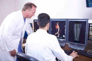
A CT uses x-rays to look inside the body to provide detailed anatomy. Sometimes there can be other processes going on inside the abdomen that may block the flow in the testicular vein. The CT scan is also performed to accurately measure the size of the testicular veins. Our specialist routinely orders this exam prior to treatment to allow for more precise placement of the coils to prevent migration or complication.7,9
Is embolization possible?
Embolization works by navigating a catheter into the testicular vein and sealing it. Whether — and how — that can be done depends on anatomy the ultrasound never sees: the course, size and number of the testicular veins, whether there are duplicated or collateral channels, and how the vein joins the renal vein or vena cava.7 Mapping this in advance is what allows the coils to be placed precisely, and it is why the CT is obtained before treatment rather than discovered during it.
Ruling out other causes
Most varicoceles are primary — the valves in the testicular vein fail, blood pools, and the veins enlarge.2 But a varicocele can also be secondary: a sign that something higher up is obstructing the vein.6,9 The CT is what rules this in or out, including:
- Vascular compression — the left renal vein can be compressed between arteries (the "nutcracker" phenomenon), raising pressure that backs up into the testicular vein.5
- A mass or tumor in the abdomen, retroperitoneum, or kidney pressing on or invading the vein.6
- Other obstructing processes inside the abdomen that block flow in the testicular vein.
Why this matters. Treating a secondary varicocele without identifying what's causing it means the real problem goes undiagnosed. Findings that raise suspicion — a right-sided varicocele on its own, a sudden onset, or one that doesn't decompress when lying down — are exactly what this scan is designed to catch — published workup guidance advises investigating with ultrasound or CT before repairing the varicocele.9
What to expect from your evaluation
Exam, then ultrasound, then CT — and finally a plan built on all three, reviewed with you by the doctor who will perform the procedure.
What is varicocele embolization?
A non-surgical, outpatient, minimally invasive technique using X-ray guidance to place tiny coils and embolic fluid in the abnormal veins, closing them down.
Varicocele can be treated by an embolization procedure or surgery. Embolization is a non-surgical, outpatient, minimally invasive technique that uses x-ray guidance to place tiny coils and embolic fluid in the abnormal blood vessels causing them to close down.7
Am I a candidate for varicocele embolization?
Treatment is offered for males who have:
- Fertility problems (problems fathering a child)8
- Pain
- Boys with a smaller testicle; at a higher risk for fertility problems when they are older8
How is non-surgical embolization performed?
- You will receive twilight sedation to help you relax
- Our specialist numbs the skin on the neck or groin
- A tiny catheter is placed
- Foam and small coil plugs placed into the vein
- The catheter is removed
- A Band-Aid is applied
The same doctor reads your imaging and performs your procedure
Being dual board certified in diagnostic imaging and interventional radiology means the ultrasound and CT aren't handed off to someone else — he reads them, and he treats what he sees.
Some skills can't be taught — only sharpened
Dual board certified in imaging — so the study can be done in our office and interpreted directly, not read off someone else’s report.
Routine pre-treatment CT allows precise coil placement to prevent migration or complication — and catches causes an ultrasound can’t see.
Request an Appointment
Please note that although we strive to protect and secure our online communications, and use the security measures detailed in our Privacy Policy to protect your information, no data transmitted over the Internet can be guaranteed to be completely secure and no security measures are perfect or impenetrable. If you would like to transmit sensitive information to us, please contact us, without including the sensitive information, to arrange a more secure means of communication. By submitting this form you consent to receive text messages from CVI at the number provided. Msg & data rates may apply. Msg frequency varies. Unsubscribe at any time by replying STOP.
Who evaluates and treats you?
At Varicocele Doctor, your evaluation and treatment are performed by Dr. Atabak Allaei — a dual board-certified Interventional Radiologist, the only specialty in medicine specifically trained to perform embolization procedures.
It matters who reads the images. A varicocele workup is a set of flat pictures of a three-dimensional venous system, and the physician has to reconstruct that anatomy mentally before a catheter ever moves. That spatial skill comes easily to very few, and experience alone can’t manufacture it — it can only sharpen it in a physician who already has it.
Atabak Allaei, MD
Dual board certified in diagnostic imaging and interventional radiology, Dr. Allaei can perform and interpret your scrotal ultrasound himself and uses a pre-treatment CT to plan precise coil placement. Over 2,000 complex embolizations across more than 10,000 image-guided procedures. All appointments are conducted by the board-certified doctor — not assistants or non-physician providers. Consultations by telehealth or in person in Los Angeles, Orange County, and San Diego.
Diagnosing a varicocele: common questions
How is a varicocele diagnosed?
Diagnosis usually involves three steps. First, a physical examination of the testicles in standing or lying positions, since a varicocele cannot always be seen. Second, a scrotal ultrasound — the best way to see inside the scrotum and determine severity — performed with the Valsalva maneuver, where you bear down to see if the veins enlarge. If the veins meet a certain size criterion the diagnosis of a varicocele can be made. Third, a CT scan to accurately measure the testicular veins, determine whether embolization is possible, and rule out other vascular and tumor causes.
Can a physical exam alone diagnose a varicocele?
Not reliably. A varicocele cannot always be seen or felt, so an exam can miss smaller ones. Just as importantly, an exam cannot tell you what else might be going on: other conditions — including a tumor, an infection, or a cyst such as a spermatocele or hydrocele — can produce similar scrotal swelling, heaviness, or discomfort. That is why imaging is used to confirm what is actually present rather than treating based on feel alone.
Why is scrotal ultrasound the best test?
Ultrasound uses high-frequency sound waves to create an image inside your body, and because the scrotum is located on the outside of the body it is an ideal test — there is no radiation and nothing to prepare for. It is the best way to see inside the scrotum, confirm the diagnosis, and determine how severe the varicocele is. The study is done with the Valsalva maneuver: the technologist may have you bear down like you are going to the bathroom to see if the veins enlarge. If the veins meet a certain size criterion the diagnosis can be made, and sometimes the abnormal flow can also be visualized.
Why is a CT scan needed before treatment?
A CT uses X-rays to look inside the body and provide detailed anatomy, and it answers two questions an ultrasound cannot. First, it shows whether embolization is possible and how it should be planned — accurately measuring the size of the testicular veins and mapping the anatomy, which allows for more precise placement of the coils to prevent migration or complication. Second, it rules out other causes: there can be other processes inside the abdomen that block flow in the testicular vein, including vascular compression and tumors or masses. Our specialist routinely orders this exam prior to treatment.
What else can cause a varicocele?
Most varicoceles are primary — caused by faulty valves in the testicular vein. But a varicocele can also be secondary, meaning it is a symptom of something else obstructing the vein higher up: compression of the renal vein between arteries, or a mass or tumor in the abdomen or kidney pressing on the vein. Features that raise suspicion include a varicocele on the right side alone, one that appears suddenly, or one that does not decompress when lying down. This is precisely what the CT is designed to catch, because treating the varicocele without identifying an underlying cause would leave the real problem undiagnosed.
Do I need a new ultrasound if I already had one?
Typically by the time patients come to our office they have already been diagnosed with a varicocele. If you do not have an ultrasound our specialist will either order one at a location close to you or perform this study at our office — our doctor is dual board certified and can perform ultrasound studies himself. That also means he interprets the images directly rather than relying solely on an outside report.
Am I a candidate for varicocele embolization?
Treatment is offered for males who have fertility problems (problems fathering a child), pain, or — in boys — a smaller testicle on the affected side, who are at higher risk for fertility problems when they are older. A consultation reviews your imaging, labs and history to determine whether you are a candidate for the procedure and the outcomes you can expect.
Who performs my evaluation and treatment?
All appointments are conducted by our board-certified doctor and not assistants or non-physician providers. He is dual board certified in diagnostic imaging and interventional radiology, which means the same physician who reads your ultrasound and CT is the one who performs your embolization — with over 2,000 complex embolizations across more than 10,000 image-guided procedures. Appointments are available via online video telehealth or in person in Los Angeles, Orange County or San Diego.
- Cimador M, Di Pace MR, Peritore M, Sergio M, Castagnetti M, De Grazia E. The role of Doppler ultrasonography in determining the proper surgical approach to the management of varicocele in children and adolescents. BJU Int. 2006;97(6):1291-1297.
- Stanford Health Care. Varicocele.https://stanfordhealthcare.org/medical-conditions/mens-health/varicocele.html
- Belay RE, Huang GO, Shen JK, Ko EY. Diagnosis of clinical and subclinical varicocele: how has it evolved? Asian J Androl. 2016;18(2):182-185. doi:10.4103/1008-682X.169991. PMID: 26780869.
- Srivastav S, Jamil RT, Zeltser R. Valsalva Maneuver. In: StatPearls [Internet]. Treasure Island (FL): StatPearls Publishing.https://www.ncbi.nlm.nih.gov/books/NBK537248/
- Kurklinsky AK, Rooke TW. Nutcracker phenomenon and nutcracker syndrome. Mayo Clin Proc. 2010;85(6):552-559. doi:10.4065/mcp.2009.0586. PMID: 20511485.
- Robson J, Wolstenhulme S, Knapp P. Is there a co-association between renal or retroperitoneal tumours and scrotal varicoceles? A systematic review. Ultrasound. 2012;20(4):182-191. doi:10.1258/ult.2012.012020.
- Halpern J, Mittal S, Pereira K, Bhatia S, Ramasamy R. Percutaneous embolization of varicocele: technique, indications, relative contraindications, and complications. Asian J Androl. 2016;18(2):234-238. doi:10.4103/1008-682X.169985. PMID: 26658060.
- Practice Committee of the American Society for Reproductive Medicine; Society for Male Reproduction and Urology. Report on varicocele and infertility: a committee opinion. Fertil Steril. 2014;102(6):1556-1560. doi:10.1016/j.fertnstert.2014.10.007. PMID: 25458620.
- Kim ED, et al. Varicocele Workup: Laboratory Studies, Imaging Studies. Medscape Drugs & Diseases.https://emedicine.medscape.com/article/438591-workup
The above information explains what is involved and the possible risks. It is not meant to be a substitute for informed discussion between you and your doctor, but can act as a starting point for such a discussion.
Explore next steps
Varicocele Embolization
The procedure itself, step by step.
Varicocele Symptoms
Pain, fertility problems and what they mean.
Hydrocele Treatment
Fluid around the testicle — a different problem.
Comparison Chart
Embolization vs. surgical options.
Clinical Evidence
What the research shows.
Meet Our Specialist
The physician who evaluates and treats you.
We are here to help
Request an appointment to meet with our varicocele specialist, who will review your imaging, labs and history to determine if you are a candidate for the procedure, and the outcomes you can expect. Each person is an individual and should discuss the potential risks and benefits of embolization and other treatments with our doctor to decide which option is best. Appointments are available via an online video telehealth platform or in person in Los Angeles, Orange County or San Diego.